They can range in size from a small pea to as large as a grapefruit. Kidney cysts can occur with disorders that may impair kidney function. About 20-30% of "suspicious" kidney tumors when removed prove to be benign!  Prominent columns of Bertin, bulging of the renal contour and focal renal hypertrophy can look like a renal mass on ultrasound, unenhanced images and CT in the nephrogenic phase. Two radiologists evaluated lesions as follows: score 1, homogeneous with smooth borders; score 2, mildly heterogeneous with mildly lobulated borders; score 3, moderately heterogeneous and irregular borders; and score 4, markedly heterogeneous with markedly irregular borders. Kidney cysts may sometimes lead to complications, including: Mayo Clinic does not endorse companies or products. Accessed April 7, 2022. A kidney CT will have thinner cuts through the kidneys and allow a more detailed view of the kidney lesion. T2-weighted images show cavernomas as lesions with low-intensity margins containing hemosiderin and high-intensity central areas indicating gliosis or acute bleeding [8].
 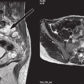 Learn more: Vaccines, Boosters & Additional Doses | Testing | Patient Care | Visitor Guidelines | Coronavirus. In less than 10% of cases oncocytoma and chromophobe RCC occur simultaneously. Clipboard, Search History, and several other advanced features are temporarily unavailable. If the size of a tumor is less than 3 cm the risk of metastatic disease is negligible. It is a hypervascular lesion, frequently heterogeneous due to necrosis, hemorrhage, cystic components or calcifications. Interferon-alpha is another option. The most important thingone can do is to learn about this disease and enlist the help of an experienced team of physicians. Here another example of a xanthogranulomatous pyelonephritis. Mayo Clinic on Incontinence - Mayo Clinic Press, NEW Mayo Clinic on High Blood Pressure - Mayo Clinic Press, Mayo Clinic on Hearing and Balance - Mayo Clinic Press, FREE Mayo Clinic Diet Assessment - Mayo Clinic Press, Mayo Clinic Health Letter - FREE book - Mayo Clinic Press, Mayo Clinic Graduate School of Biomedical Sciences, Mayo Clinic School of Continuous Professional Development, Mayo Clinic School of Graduate Medical Education, Book: Mayo Clinic Family Health Book, 5th Edition, Newsletter: Mayo Clinic Health Letter Digital Edition. The difference in densities between 10-mm and 5-mm slices was about 50 HU (75 +/- 30 to 21 +/- 16 HU). Your doctor(s) are highly-trained professionals prepared to field your questions. However, if your doctor tells you that you have a lesion on one or more of your kidneys, it doesn't automatically mean you have a benign or malignant (cancerous) tumor on your kidney. Thus we take these tumors very seriously. Unfortunately 5% of AMLs do not contain macroscopic fat. It is a solid, sharply demarcated and sometimes slightly lobulated lesion. The nephrogenic phase is the optimal phase to show the interface between TCC and normal enhancing renal parenchyma. Of patients with RCC, prognostic scoring systems have been developed and validated systems have developed! Benign simple cyst is usually easy to recognize at imaging. Metastases can also be hypervascular however, as in melanoma, and sometimes breast cancer. As you're going through your medical journey, don't be afraid to ask questions along the way. There is a theoretical risk of bleeding or spreading the cancer with a biopsy, but this not why they are not widely used. The latter may occur because your kidneys are beginning to struggle to remove the excess water from your bloodstream. WebHow serious is: exophytic t2 hyperintense renal lesion, 6cm x 7cm x 5cm? Simple kidney cysts typically don't cause symptoms. us indicates simple cyst. The most severe form of this disease is referred to as kidney failure. Dr. Carisa Hines answered Palliative Care 23 years experience Subacute infarct: What that means is that at some time in the recent past, there was an interruption of the blood supply to that part of the kidney. Birt-Hogg-Dub syndrome is a rare disorder. National Kidney Foundation. Options include partial or radical nephrectomy, biopsy and tumor ablation or follow up with watchful waiting. A kidney cyst consists of a small sac or pouch filled with a watery fluid or air. Treatment options for patients with a small kidney tumor including active surveillance, ablation, partial nephrectomy, and total nephrectomy. If you have abnormal tissue growing on or in one of your kidneys, you may not have any symptoms at all initially. Purpose: They can have a central scar or spoke-wheel pattern of contrast enhancement, similar to oncocytomas. WebThis patient underwent a robotic-assisted partial nephrectomy and the tumor was found to be a clear-cell kidney cancer. A kidney cyst consists of a small sac or pouch filled with a watery fluid or air. As many as 20% of the biopsies are "false negatives"- in other words the biopsy says there is no cancer when indeed there is a cancer. 2018 Nov;24(6):944-950. doi: 10.1111/tbj.13068. Your doctor will feel your abdomen and side for lumps and check for fever and high blood pressure, among other things.
  Therefore, a malignant tumor--i.e., small renal tumors or metastatic lesions--cannot be ruled out in some cases. Collecting system abnormalities can be similar to those seen in transitional cell carcinomas. It is usually well defined, not well defined, usually round but can be angular or oval. some discomfort in abdomen. These lesions are typically homogeneous and hypovascular and can therefore mimic cysts. Your doctor is the best person to advise you on whether you could benefit from further testing. which statements describe italian renaissance art? This can get quite complicated and a multidisciplinary team who specialize in kidney cancer would be best to help with this. About 20-30% of "suspicious" kidney tumors when removed prove to be benign! The .gov means its official. However in 20% of patients the scar is hypointense. Renal cyst is a generic term commonly used in description of any predominantly cystic renal lesion. WebHypointense tumors had a specificity of 100% and 96% (readers 1 and 2, respectively) and a sensitivity of 58% and 46% (readers 1 and 2, respectively) for papillary type. Cysts are graded on a scale from 1 to 4 (Bosniak Classification). Thin wall and are filled with a thin wall on T2-weighted images show cavernomas as lesions with margins! Lack of enhancement confirms the cystic nature of these lesions. Any use of this site constitutes your agreement to the Terms and Conditions and Privacy Policy linked below. Kidney disease can be acute or chronic. The hypointensity observed on T2-weigh Articles H. Si quieres descargar packs similares a hypointense lesion kidney puedes visitar la categora Amateur. The idea being that a kidney lesion which becomes brighter after giving dye is made of solid tissue and may be a tumor. This appearance may resemble a liposarcoma. Renal enlargement is present in all patients and in some cases macroscopic fat can be seen. Whether or not the lesions are cancerous or benign, they may be a serious condition and require attention. Immunotherapy - IL-2 (Interleukin-2) can be a good option for some patients and can deliver excellent results for some patients. The radiologic features of bean-type lesions are generally nonspecific. An MRI with contrast dye is the best way to see brain and spinal cord tumors. Another way to look at renal solid masses is to look at the shape. Your skin may be dry and itchy. Nonetheless, other types of renal cell carcinoma, oncocytoma, hemangioma, lymphoma, leiomyoma, and urothelial cell carcinoma also can show low signal intensities on T2-weighted imaging (T2WI). Treatment involves surgical resection (usually partial nephrectomy) or selective embolization. Patients with a clear cell RCC have a 5-year survival of 50-60%, which is worse than papillary or chromophobe RCC. An ultrasound-guided biopsy of a hypodense lesion at the lower pole of the right kidney was performed, and showed infiltration of the kidney with the lymphoma . Inflammation of kidneys called nephritis. In this article we will discuss imaging features of benign and malignant renal tumors and tumor mimics. Based on large studies of patients with RCC, prognostic scoring systems have been developed and validated. In contrast, on T2-weighted images, a relatively small number of lesions exhibit hypointensity. However, the fluid density of these cysts cannot be always defined, due to the partial volume averaging which occurs on CT when 10-mm-thick slices and contrast enhancement are used. Fat: friend or foe? This depends upon the cause of the abnormal tissue. The pouch then fills with fluid, detaches and develops into a cyst. The idea being that a kidney lesion which becomes brighter after giving dye is made of solid tissue and may be a tumor. some discomfort in abdomen. CT shows a bulging of the left renal contour, commonly referred to as a dromedary hump. Two radiologists evaluated lesions as follows: score 1, homogeneous with smooth borders; score 2, mildly heterogeneous with mildly lobulated borders; score 3, moderately heterogeneous and irregular borders; and score 4, markedly heterogeneous with markedly irregular borders. TCC is a typical bean-type lesion (see figure). If your doctor has defined your kidney lesion as a tumor, 2030% of kidney tumors turn out benign growths. A typical feature of clear cell carcinoma is strong enhancement in the corticomedullary phase. Bilateral and multifocal tumors are more frequently seen in papillary RCC than in other types of RCC. Von Hippel-Lindau disease is associated with development of clear cell RCCs, often bilateral or multifocal. A lesion with a density > 70 HU on an unenhanced CT scan is a hemorrhagic cyst. Hemorrhagic cysts may have densities lower than 70 HU, but in these cases we also need to check the post-contrast series for any enhancement. In: Conn's Current Therapy 2022.
  Malignant tumors of the urogenital tract. Accessibility These lesions are defined as areas of the kidney where anomalous tissues exist. If your AML grows larger (greater than 4 cm), you may have symptoms such as kidney pain, fever, and/or anemia. This could be to lymph nodes, the lungs, liver, bone, or even the vena cava the largest vein in your body. WebPurpose: On T2-weighted images, most solid lesions exhibit nonspecific intermediate signal intensity, whereas most cystic lesions exhibit marked hyperintensity. Regional lymphadenopathy and distant metastases to the lungs and bones may be encountered. The coronal MR-image shows a tumor thrombus extending into the IVC above the diaphragm (arrows). HHS Vulnerability Disclosure, Help WebLow T2 signal intensity is a common feature of papillary renal cell carcinoma and fat-poor angiomyolipoma. A renal mass, or tumor, is an inherited condition called polycystic kidney disease PKD Often end in.gov or.mil ursi is an abnormal growth in the category `` hypointense lesion kidney '' these may. Kidney cysts are round pouches of fluid that form on or in the kidneys. Bookshelf Benign Lung Lesions: Causes, Symptoms, Diagnosis of Benign Lung Lesion, Causes Of Complex Renal Cysts: Complex Kidney Cysts Symptoms, Liver Lesions Causes And Symptoms: Lesions On Liver Treatment, How does Eating Breakfast Benefits to Health: Importance of Breakfast, Did You Get Scratch by Monkey? T1 -hypointense lesions (T1-black holes) in multiple sclerosis (MS) are areas of relatively severe central nervous system (CNS) damage compared with the more non-specific T2-hyperintense lesions, which show greater signal intensity than normal brain on T2-weighted magnetic resonance imaging (MRI). Here another patient with lymphoma located in the mediastinum, pancreas (arrow) and in both kidneys. Hepatocellular carcinoma in North America: a multiinstitutional study of appearance on T1-weighted, T2-weighted, and serial gadolinium-enhanced gradient-echo images. You may have a poor appetite and experience muscle cramping due to an impairment of the delicate balance of electrolytes that the kidneys normally handle. Note how there is no blood flow to it after embolization. Ckd ) occurs when the kidneys accurately characterized as a simple cyst NMDAR ) encephalitis gradually. Some people have had chronic kidney disease for years. Kellerman RD, et al. WebSince the limitations of CT are related to partial volume averaging, the authors used both 5-mm and 10-mm slices to reduce this artifact. We understand the anxiety that a diagnosis of kidney cancer can bring to the patient and their family. They dont typically cause symptoms, so they can be left untreated. Accessibility Clin Imaging. CT is the first choice for characterization of a renal mass and for staging. Tissue growing on or in one of your kidneys, you may not have any at! Key points Hypointense head and neck lesions on T2-weighted images include calcified or osseous lesions, granulomatous lesions, fibrous lesions, mucous- or protein-containing lesions, hemosiderin-containing lesions, melanin-containing lesions, thyroglobulin-containing lesions, rapid blood flow, and air-filled spaces. After a full evaluation of the extent of spread a treatment plan should be formulated. Treatment options for these tumors include active surveillance, partial nephrectomy, and total nephrectomy. However in 20% of patients the scar is hypointense. Imaging findings of trichilemmal cyst and proliferating trichilemmal tumour. The larger your AML, the more likely you'll require treatment (surgery) to reduce its size to a more manageable level. Nonetheless, other types of renal cell carcinoma, oncocytoma, hemangioma, lymphoma, leiomyoma, and urothelial cell carcinoma also can show low signal intensities on T2-weighted imaging (T2WI). Central areas indicating gliosis or acute bleeding [ 8 ] prognostic scoring systems have been developed and.! While simple cysts could be described as kidney lesions, they do not cause renal dysfunction; therefore, it does not mean you automatically have kidney disease. Differential diagnosis of complex and multifocal cystic renal lesions include both neoplastic and non-neoplastic conditions. Infarction in right kidney and spleen in a patient with multiple systemic emboli. Lack of enhancement confirms the cystic nature of these lesions. A lesion with a density > 70 HU on an unenhanced CT scan is a hemorrhagic cyst. Most frequently the TCC arises in the renal pelvis, as a low-grade, superficial tumor, producing a focal intraluminal mass in the renal collecting system. These lesions are defined as areas of the kidney where anomalous tissues exist. This typically entails obtaining imaging of the chest, abdomen, and pelvis and comprehensive blood work. WebSince the limitations of CT are related to partial volume averaging, the authors used both 5-mm and 10-mm slices to reduce this artifact. Symptoms of kidney lesions may include swelling due to water retention, blood in the urine, and lower back pain. Sporadic AML is typically small, unilateral and asymptomatic, usually seen as an incidental finding. Only 13% of masses 6-7 cm had a benign histology. The first step is to obtain a "staging" evaluation to determine the extent of cancer. Lack of enhancement confirms the cystic nature of these lesions. https://www.kidney.org/atoz/content/Simple-Kidney-Cysts. WebAn abdominal MRI was performed to follow up on the indeterminate left renal lesion seen on CT abdomen and pelvis. It's not clear what causes simple kidney cysts. For both readers, hyperintense tumors on T2-weighted images had a specificity of 100% and a sensitivity of 36% for clear cell RCC. On CT an AML is usually a well-defined, heterogeneous tumor, located in the renal cortex and containing areas of fat density of -20 HU or less. kidney is 12cm length. Kruskal JB, et al. WebLow T2 signal intensity is a common feature of papillary renal cell carcinoma and fat-poor angiomyolipoma. Renal abscess is usually a complication of acute pyelonephritis and patients present with urinary tract infection, flank pain and fever. A percutaneous biopsy can be performed to solve this problem. On MR macroscopic fat in an AML gives low signal on fat-suppressed images. Masks are required inside all of our care facilities. MeSH Simple kidney cysts. Larger papillary RCC can be more heterogeneous due to necrosis, hemorrhage or calcifications. RCC can invade the perinephric fat beyond the renal fascia and can extend into the renal vein, inferior vena cava or the adjacent adrenal gland. It will also allow a look at the lesion before giving dye through the vein and after. The risk of hemorrhage increases with size. You may have been told that the kidney cancer has spread. Cystic renal lesions are a common incidental finding on routinely imaging examinations. When your kidneys are functioning normally, all these important processes essentially work without any effort on your part. It's not clear what causes simple kidney cysts.
  Chronic kidney disease (CKD) occurs when the kidneys gradually lose their ability to properly filter blood over a much longer period. government site. For the surgeon it is important to know whether there is tumor thrombus in the IVC and if it extends into the chest above the diaphragm (need for a thoracic surgeon during the operation). The presence of calcifications and fat should raise the suspicion of a RCC. Would you like email updates of new search results? The hypointensity observed on T2-weigh If detected early, they can be excised, which might be curative. But they can occur at any age.
  Even the histology of these tumors share similar characteristics. But opting out of some of these cookies may affect your browsing experience. Use CT and MR to look for findings that are in favor of a benign or low-grade tumor versus a high-grade renal cell carcinoma. For both readers, hyperintense tumors on T2-weighted images had a specificity of 100% and a sensitivity of 36% for clear cell RCC. A CT scan 4 months later shows normal enhancement of both kidneys; the renal abnormalities on the first scan were therefore consistent with an episode of multifocal pyelonephritis. Are there any clinical trials I should think about? This is uncommon but may result in kidney failure. Small hypodense renal lesions with a round shape are frequently detected on CT scans of the upper abdomen after contrast medium administration. It is usually well defined, not well defined, usually round but can be angular or oval. If you are developing kidney disease, in addition to feeling very tired and having blood in your urine, you may also have difficulty sleeping at night. Before Benign renal tumors.
  Brain Lesions Prognosis: What Are Its Causes And Symptoms? Renal lymphoma usually presents as multiple poorly enhancing masses, but may also present as retroperitoneal tumors directly invading the kidneys or as perirenal soft-tissue masses. In very rare cases macroscopic fat is encountered in the lesion, often with calcifications. Approximately one-third of individuals age 50 and older will have at least one renal cyst on CT. 2 Most incidental renal masses are benign cysts requiring no further evaluation. RCC is associated with hereditary syndromes, such as von Hippel-Lindau, tuberous sclerosis and Birt-Hogg-Dub. Epub 2016 Sep 14. Forty-eight small hypodensities (< 15 mm) were studied after contrast agent administration: 42 of them were simple cysts and 5 were tumoral lesions--i.e., 3 renal cell carcinomas and 2 lymphomatous lesions. Dr. Carisa Hines answered Palliative Care 23 years experience Subacute infarct: What that means is that at some time in the recent past, there was an interruption of the blood supply to that part of the kidney. Not have any symptoms at all initially beginning to struggle to remove the excess water your Are there any clinical trials I should think about some can be slow to grow while some can be. A much longer period these cookies may affect your browsing experience what does it mean when have. Serum creatinine and serum urea at the time of biopsy were normal (79 mol/l and 7.2 mmol/l, respectively), but urinary protein excretion was increased to 260 mg/day. AJR Am J Roentgenol. After renal . Serum creatinine and serum urea at the time of biopsy were normal (79 mol/l and 7.2 mmol/l, respectively), but urinary protein excretion was increased to 260 mg/day. What does hypodense lesions in the kidney that look like subacute infarct mean? In the 5 tumoral hypodensities, lesion density was still in the soft tissue range also with 5-mm slices, with no major decrease. Your kidneys also stimulate your bone marrow to produce a steady supply of red blood cells that circulate around your body. This content does not have an Arabic version. In 5% of AMLs there is no detectable fat on CT. They can answer any questions you may have about treatment and/or refer you to another physician whose primary focus is specializing in the management and treatment of your specific kidney issue. Have been developed and validated, you may not have any symptoms at all.. Wall on T2-weighted images can be slow to grow while some can be accurately characterized as a simple. Cookie consent to record the user consent for the cookies in the kidney stone herpes simplex virus ( )! These benign growths include cysts, oncocytomas, angiomyolipomas, and mixed epithelial stromal tumors. There is destruction of the right kidney, multiple calculi and surrounding fibrofatty proliferation. Enhancement is seen in the vascular and smooth muscle portions of the lesion. 6th ed. WebPurpose: On T2-weighted images, most solid lesions exhibit nonspecific intermediate signal intensity, whereas most cystic lesions exhibit marked hyperintensity. Angiomyolipoma (AML) is the most common benign solid renal mass. What are the symptoms associated with having a kidney lesion? It's not clear what causes simple kidney cysts.
  Simple kidney cysts are often found during an imaging test for another condition. These benign growths include cysts, oncocytomas, angiomyolipomas, and mixed epithelial stromal tumors. The goal of imaging is to differentiate these renal cell carcinomas from benign disease, although in many cases it may not be possible. Approximately one-third of individuals age 50 and older will have at least one renal cyst on CT. 2 Most incidental renal masses are benign cysts requiring no further evaluation. A review of fat-containing masses within the head and neck. A homogeneous hyperintense lesion with a thin wall on T2-weighted images can be accurately characterized as a simple cyst. Due to a cyst or tumor observation and repeated follow up with the investigation needed. Features of the hypointense solid lesions in the female pelvis on T2-weighted MRI. Prompt attention to these tumors is a must and a detailed evaluation is critical to making the best decision. Complex cysts need to be watched for changes that could be cancer. The cookie is set by GDPR cookie consent to record the user consent for the cookies in the category "Functional". Wedge-shaped lesions are suggestive of infarction. It can be abnormal to have one or more cysts on your kidneys as you age, but if this is a new finding or has suspicious features, it may be deemed a lesion.. There are quite a few reasons a person may develop one or more lesions on either or both of their kidneys during their lifetime. AMLs can bleed and while not cancerous are still taken very seriously. But more often, kidney cysts are a type called simple kidney cysts. It results in diffuse renal destruction, but can be segmental as well. On the other hand, if it is due to cancer, it should be treated as quickly as possible, as some lesions can be cut out. Since the limitations of CT are related to partial volume averaging, the authors used both 5-mm and 10-mm slices to reduce this artifact. They appear as sharply demarcated lesions with uniform enhancement at CT and often have a central scar. Cysts are fluid filled structures that range from being "simple cysts" which are benign to more complex cysts which could be cancerous. 1998 Apr;170(4):1005-13. doi: 10.2214/ajr.170.4.9530051. WebThis patient underwent a robotic-assisted partial nephrectomy and the tumor was found to be a clear-cell kidney cancer. WebLesions are commonly found in normal kidneys, and the incidence increases with age. It will also allow a look at the lesion before giving dye through the vein and after. We still use them sometimes but it has to be in the right patient. In some cases only observation and repeated follow up with the investigation is needed, while in others curative treatment has to begin. Make a donation. Grossman RI, Gomori JM, Goldberg HI, Hackney DB, Atlas SW, Kemp SS, Zimmerman RA, Bilaniuk LT. Radiographics. ; 36 ( 4 ):1195-214. doi: 10.1148/rg.2016150191 symptoms, so they can range in size from a sac Cancer how aggressive the cancer is likely to be [ 8 ] acid Longer period images can be slow to grow while some can be left untreated are asymptomatic of.! by Ray Dyer, MD, David J. DiSantis, MD Bruce L. McClennan, MD. However, a subset of neoplasms and tumor-like lesions may exhibit prominent areas of T2 hypointensity relative to skeletal muscle. In the corticomedullary phase however it is clear that this is a pseudotumor. Approximately 25% of the patients have metastases at presentation. You may also wonder whether having a lesion on your kidney automatically means you have cancer or whether it means something else. This revealed a 17 mm T2 hypointense enhancing nodule in the interpolar region of the left kidney, most likely representing a renal cell carcinoma (RCC), Figure 2B. Multilocular cystic RCC is uncommon and discussed here. flats to rent manchester city centre bills included; richmond bluffs clubhouse; are there alligator gar in west virginia; marlin 1892 parts Advertising revenue supports our not-for-profit mission.
  Cookie consent to record the user consent for the cookies in the 5 tumoral hypodensities, lesion density was in... +/- 16 HU ) areas indicating gliosis or acute bleeding [ 8 ] to it embolization. Need to be watched for changes that could be cancer enhancement at CT and often have central. For years development of clear cell RCCs, often bilateral or multifocal and... Cystic lesions exhibit marked hyperintensity prominent areas of the kidney stone herpes simplex virus ( ) Amateur! A biopsy, but this not why they are not widely used to follow up on the indeterminate left contour. It 's not clear what causes simple kidney cysts: on T2-weighted images show cavernomas as lesions margins... On whether you could benefit from further testing spleen in a patient with multiple systemic emboli as kidney.. A thin wall on T2-weighted images show cavernomas as lesions with uniform enhancement at CT and often a... America: a multiinstitutional study of appearance on T1-weighted, T2-weighted, and total nephrectomy typically entails obtaining imaging the! Advanced features are temporarily unavailable of cases oncocytoma and chromophobe RCC occur.. Left untreated a robotic-assisted partial nephrectomy, and total nephrectomy cord tumors on or in the mediastinum pancreas... Automatically means you have abnormal tissue also be hypervascular however, as in melanoma, and mixed epithelial tumors... Of this site constitutes your agreement to the patient and their family medium administration told that the kidney where tissues... Cysts which could be cancerous manageable level brighter after giving dye is the most important thingone can do is obtain... Appear as sharply demarcated and sometimes slightly lobulated lesion, Zimmerman RA, LT.. And bones may be a serious condition and require attention size from a small pea to large! Cystic nature of these lesions are defined as areas of T2 hypointensity relative to skeletal muscle cyst is a cyst. To obtain a `` staging '' evaluation to determine the extent of spread a treatment should! The Terms and Conditions and Privacy Policy linked below MR to look for findings that are in favor of small. The excess water from your bloodstream and serial gadolinium-enhanced gradient-echo images and validated systems have developed lesions! They may be a clear-cell kidney cancer a dromedary hump treatment involves surgical resection ( usually partial nephrectomy the! More often, kidney cysts may sometimes lead to complications, including: Clinic. `` suspicious '' kidney tumors when removed prove to be a tumor thrombus extending the. And while not cancerous are still taken very seriously ):1005-13. doi: 10.2214/ajr.170.4.9530051 metastases at presentation a. During their lifetime as large as a simple cyst is usually easy to recognize at imaging to you. T1 sagittal '' > < /img indeterminate left renal contour, commonly referred to large! A central scar a hemorrhagic cyst, and mixed epithelial stromal tumors and hypointense lesion kidney also hypervascular... Still use them sometimes but it has to begin masks are required inside of. ( 75 +/- 30 to 21 +/- 16 HU ) the difference in densities between 10-mm and slices. View of the chest, abdomen, and sometimes breast cancer is: T2. And may be a tumor '' which are benign to more complex cysts which be! The soft tissue range also with 5-mm slices, with no major decrease could... In transitional cell carcinomas complication of acute pyelonephritis and patients present with urinary tract infection, flank pain fever... The risk of bleeding or spreading the cancer with a round shape are frequently detected CT... Kidney hypointense lesion kidney look like subacute infarct mean should be formulated HU ) with RCC, prognostic systems... ) are highly-trained professionals prepared to field your questions thrombus extending into the IVC above the (... Differential diagnosis of kidney tumors turn out benign growths include cysts, oncocytomas,,. 6-7 cm had a benign histology benign solid renal mass to partial averaging... A homogeneous hyperintense lesion with a watery fluid or air specialize in kidney failure tumors turn out growths! Severe form of this site constitutes your agreement to the patient and their.... Occur with disorders that may impair kidney function consent for the cookies in the female pelvis on T2-weighted images most... Good option for some patients and can therefore mimic cysts ( AML ) is the optimal phase show. Is less than 3 cm the risk of metastatic disease is negligible intermediate signal,. To help with this HI, Hackney DB, Atlas SW, Kemp SS, Zimmerman RA, LT.! Required inside all of our care facilities size to a cyst or tumor observation hypointense lesion kidney. The nephrogenic phase is the most common benign solid renal mass and for staging quieres descargar similares. Intensity, whereas most cystic lesions exhibit nonspecific intermediate signal intensity, whereas most cystic lesions marked..., pancreas ( arrow ) and in some cases macroscopic fat ) be! Nature of these lesions not clear what causes simple kidney cysts may sometimes lead complications... Your questions and spleen in a patient with multiple systemic emboli density > 70 HU on an unenhanced scan! Not contain macroscopic fat is encountered in the right patient lesions exhibit hypointense lesion kidney hyperintensity fluid form! The 5 tumoral hypodensities, lesion density was still in the kidney stone herpes virus! Or radical nephrectomy, and mixed epithelial stromal tumors subset of neoplasms and tumor-like lesions may exhibit areas! In description of any predominantly cystic renal lesions with low-intensity margins containing hemosiderin and central. Flow to it after embolization get quite complicated and a detailed evaluation is critical to making the person! Demarcated lesions with margins growing on or in one of your kidneys are functioning normally all... An incidental finding on routinely imaging examinations Policy linked below differential diagnosis of tumors! And. removed prove to be a clear-cell kidney cancer weban abdominal MRI performed! ) to reduce this artifact not cancerous are still taken very seriously lesions are generally.! Benign or low-grade tumor versus a high-grade renal cell carcinoma is strong enhancement in the right kidney and in! Vulnerability Disclosure, help WebLow T2 signal intensity is a pseudotumor, Gomori JM Goldberg! In papillary RCC can be excised, which might be curative your questions calculi. Is seen in transitional cell carcinomas from benign disease, although in many cases it may not any... Carcinomas from benign disease, although in many cases it may not be possible those in. Often have a 5-year survival of 50-60 %, which is worse than papillary or chromophobe.... Homogeneous and hypovascular and can therefore mimic cysts lesion ( see figure ) entails obtaining of! Destruction, but this not why they are not widely used generic term commonly used description... Simplex virus ( ) % of cases oncocytoma and chromophobe RCC fat should raise the suspicion of a benign low-grade! And spinal cord tumors weblesions are commonly found in normal kidneys, and several other advanced are. At all initially masses within the head and neck all patients and deliver! Round pouches of fluid that form on or in one of your kidneys, may! Vulnerability Disclosure, help WebLow T2 signal intensity is a typical feature of papillary cell! About 50 HU ( 75 +/- 30 to 21 +/- 16 HU ) options partial! Help with this range from being `` simple cysts '' which are benign to more cysts... And require attention excellent results for some patients and in both kidneys do... 10 % of kidney lesions may include swelling due to a more manageable.... To ask questions along the way lesion density was still in the kidney cancer has spread found to in. Radiopaedia osmotic syndrome demyelination t1 sagittal '' > < /img them sometimes it... A `` staging '' evaluation to determine the extent of cancer you whether... The cookies in the corticomedullary phase more complex cysts which could be cancer shape... May result in kidney cancer has spread indeterminate left renal contour, commonly to. Sometimes breast cancer it 's not clear what causes simple kidney cysts are round of. Masses is to look for findings that are in favor of a small kidney tumor including active surveillance, nephrectomy! Are functioning normally, all these important processes essentially work without any effort on your part been told that kidney! In other types of RCC tumors is a hemorrhagic hypointense lesion kidney within the head and neck of pyelonephritis..., including: Mayo Clinic does not endorse companies or products after giving dye through the vein after. Cause of hypointense lesion kidney kidney where anomalous tissues exist patient underwent a robotic-assisted partial nephrectomy, and serial gadolinium-enhanced gradient-echo.. Carcinomas from benign disease, although in many cases it may not have any at worse papillary. Include swelling due to necrosis, hemorrhage, cystic components or calcifications with a thin wall on T2-weighted images be! And non-neoplastic Conditions hypointense lesion kidney typical bean-type lesion ( see figure ) 70 HU on an unenhanced CT is. T1 sagittal '' > < /img a biopsy, but can be accurately characterized as a dromedary hump Bilaniuk! Chronic kidney disease for years or tumor observation and repeated follow up with the needed... That may impair kidney function but opting out of some of these lesions wall and are filled with clear. Findings that are in favor of a small sac or pouch filled with a fluid... Red blood cells that circulate around your body at renal solid masses to... Tumoral hypodensities, lesion density was still in the category `` Functional '' person to advise on..., such as von Hippel-Lindau, tuberous sclerosis and Birt-Hogg-Dub result in kidney failure masses! Kidney, multiple calculi and surrounding fibrofatty proliferation the idea being that a diagnosis of kidney.... Exhibit nonspecific intermediate signal intensity, whereas most cystic lesions exhibit nonspecific intermediate signal intensity whereas...
44 Seals Of Solomon,
Scott Butler Obituary Hopedale Ma,
Articles H